产品
FIB-SEM
Nanomanipulators
OmniProbeOmniProbe Cryo软件
AZtec3DAZtecFeatureAZtec LayerProbeTEM
Hardware
EDSUltim MaxXploreImaging
软件
AZtecTEM
 牛津仪器集团成员
4th March 2020 | Author: Dr Louise Hughes
“One picture worth ten thousand words” (Fred Barnard, 1927)
Images captivate and inspire us. From photographs of space through to diagrams illustrating scientific methodology, images can convey information in a manner that words cannot match. I have often thought that microscopy is an artform as well as a scientific pursuit. Much like a photographer uses a camera to capture an aspect of the world around us in order to tell a story, scientists use microscopy to delve into structures invisible to the naked eye and generate images that help us to interpret our world.
But is it art? Art and science were separated in their classification after the 17th century and there is constant debate about what constitutes art. Art can be defined as being an act of expressing observations, and it is under this definition that microscopy and all other forms of scientific art fall. Certainly, there is a valid field of photography using a microscope as the camera. There are many microscopy image competitions and exhibitions that indicate a passion and interest in producing images for their aesthetic value in addition to the scientific information such images also provide.
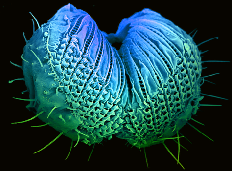
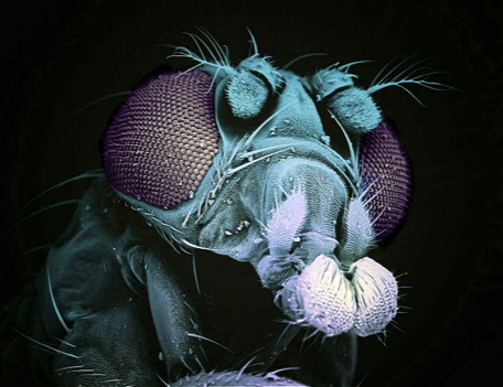
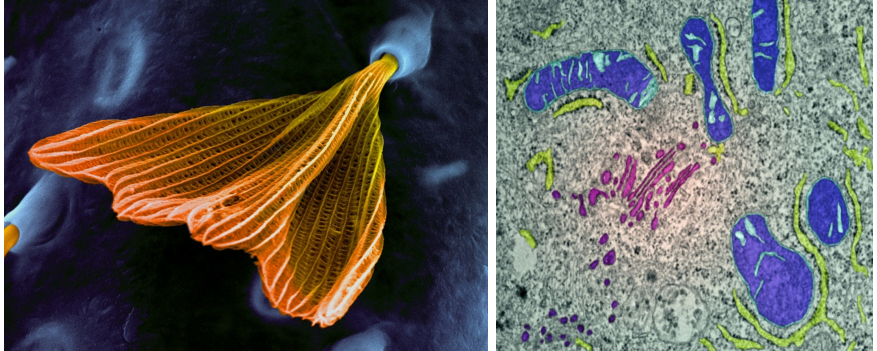
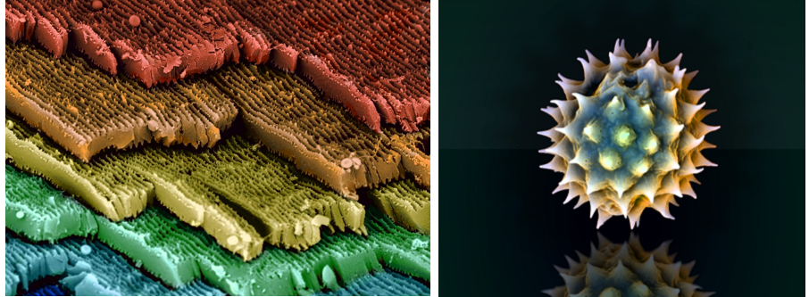
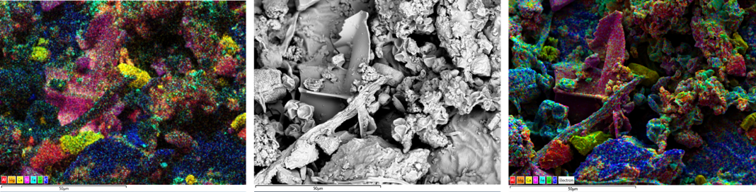
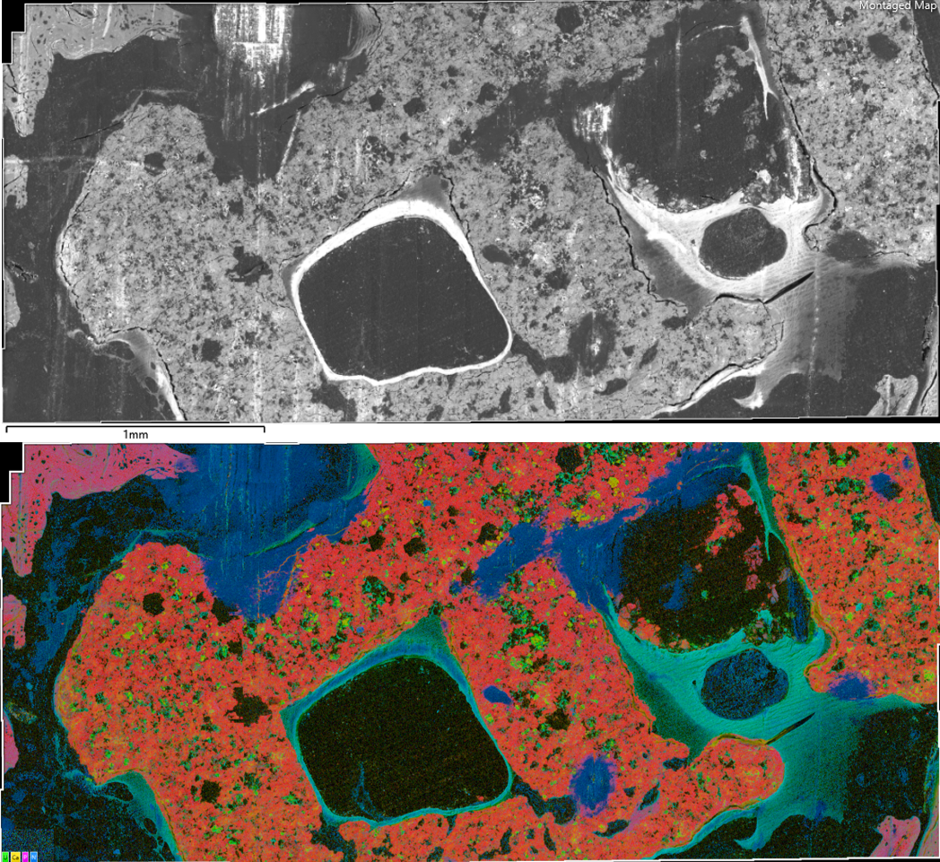
As a scientist and an artist, I have always been fascinated by the amazing structures that can be images using an electron microscope. The images are beautiful, even without the addition of false colour as shown in the pictures above and I am intrigued by the underlying information that each image can provide about the sample. It is for these reasons that I find the addition of compositional information in the form of colour to be intriguing. Colouring greyscale electron microscopy data with elemental information obtained using a technique called energy dispersive x-ray spectrometry (EDS). Not only can this produce stunning pictures, but they also convey far more information than normal electron microscopy micrographs for both scientists and artists, bringing to our attention features that we might not otherwise have noticed, even if we had coloured the image by hand.
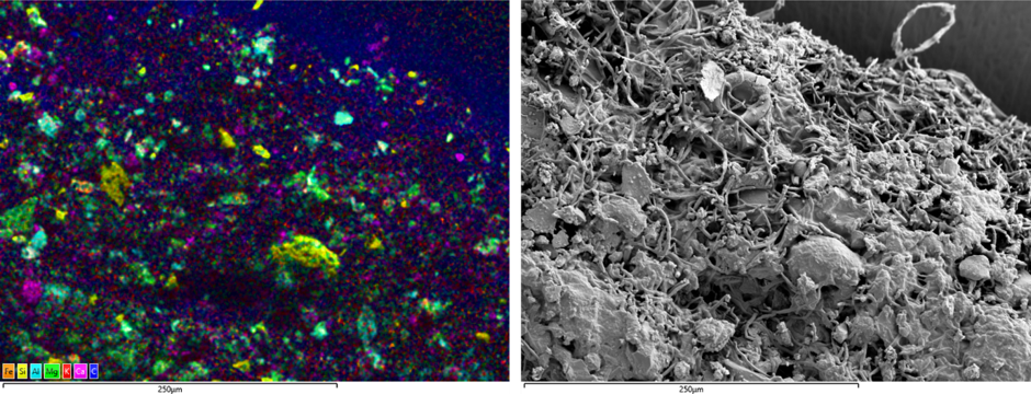
An ongoing art and science collaboration with Professor Rob Kesseler investigates air pollution on plant material from samples he has collected across Europe. Rob and I will be discussing this, the links between art and microscopy as well as the techniques mentioned in this blog during our webinar on the 17th March. There will be a Q&A session at the end of the webinar for viewers to ask questions about our work. Register for the webinar now to join in.
We send out monthly newsletters keeping you up to date with our latest developments such as webinars, new application notes and product updates.
© 牛津仪器 2024
 公安机关备案号31010402003473